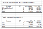
This is due to overestimation of lesions related to colposcopic changes in pregnancy, which often leads to systematic sampling despite the examiner’s experience. In our cohort, we found a prenatal biopsy rate of 45% due to suspicion of cancerous lesion. Regarding colposcopic impressions, the results are consistent with final diagnoses in 73% of cases within one degree of severity. The correlation between cytology and final diagnosis within one degree of severity in pregnant woman has been found to be 78%, with cytology having an 88% positive predictive value for high-grade lesions. The overall regression rate in our cohort was 43% (34/79). Seventy-one percent (56/79) of our cohort had a histological diagnosis at postpartum colposcopic follow up. A total of 47 biopsies and 42 conizations were performed with a rate of 53% (25/47) and 79% (33/42), respectively, of persistent high-grade lesions. Sixteen patients were only diagnosed cytologically and not histologically, of which only 3 (11%) were in the HSIL group. For patients who only underwent colposcopic evaluation (15/79), 13 histological samples were taken, 8 (62%) of which confirmed a HSIL lesion. In the 34 cytological specimens showing an LSIL/ASCUS/normal lesion, only 20 (59%) histological specimens were collected, of which 10 (50%) confirmed a high-grade lesion. Of the 27 patients whose postpartum cytology revealed an HSIL, 18 (66.7%) histological samples confirmed a high-grade lesion (either by directed biopsy or conization result) and 6 (22%) samples did not. These three cases underwent a LEEP procedure immediately postpartum due to CIN2+ extensive antenatal lesions, which was confirmed by the pathology. However, larger cohorts are required to confirm these results.Īt postpartum colposcopic follow up, all patients underwent colposcopy with cytology/histology except for three. A cervical cytology should be performed at the third trimester to identify patients at risk of CIN persistence after delivery. Parity status may have an impact on dysplasia regression during pregnancy. Conclusion: Our regression rate was high, at 43%, for high-grade cervical lesions postpartum. The presence of HSIL on third-trimester cervical cytology (OR = 0.17 95%CI = (0.04–0.72) p = 0.016) was identified as an independent predictive factor of high-grade dysplasia persistence at postpartum. Nulliparity (OR = 4.35 95%CI = (1.03–18.42) p= 0.046) was identified by multivariate analysis as an independent predictive factor of high-grade dysplasia regression. Univariate analysis revealed that parity ( p = 0.04), diabetes ( p = 0.04) and third trimester cytology ( p = 0.009) were associated with dysplasia regression. High-grade cervical lesions were diagnosed by cytology in 87% of cases (69/79) and confirmed by histology in 45% of those (31/69). Results: Between January 2000 and October 2017, 79 patients fulfilled the inclusion criteria and were analyzed.

A logistic regression model was applied to determine independent predictive factors for high-grade cervical dysplasia regression after delivery. Postpartum regression was defined cytologically or histologically by at least a one-degree reduction in severity from the antepartum diagnosis. High-grade lesions were defined either cytologically, by high squamous intraepithelial lesion/atypical squamous cells being unable to exclude HSIL (HSIL/ASC-H), or histologically, with cervical intraepithelial neoplasia (CIN) 2+ (all CIN 2 and CIN 3) during pregnancy. Methods: Pregnant patients diagnosed with high-grade lesions were identified in our tertiary hospital center. Objective: The aim of this study was to describe the evolution of high-grade cervical dysplasia during pregnancy and the postpartum period and to determine factors associated with dysplasia regression.


 0 kommentar(er)
